Mission
Neurology Networks tries to offer broad exposure to various topics that may be presented on the veterinary neurology board exam.
GNP 6
GNP 6
Q. Identify the 3 types of brain herniation (this may be up to 6 depending on your education!).
A.
1. Subfalcine: medial displacement of one cerebral hemisphere under the falx. This will damage the cingulated gyrus so is also sometimes called cingular herniation. On imaging, you may refer to this as a “midline shift”.
2. Transtentorial: displacement of the caudal aspect of the cerebral hemispheres under the tentorium cerebella. This will compress the mesencephalon, the rostral cerebellum, and the caudomedial occipital lobe and sometimes parahippocampal gyrus.
3. Cerebellar coning: displacement of the caudaoventral cerebellar vermis through the foramen magnum. This will usually include the pyramic and uvula lobules and will compress the medullary tissue at that level.
Veterinary Neuroanatomy and Clinical Neurology, 3rd edition. De Lahunta, Glass. Saunders Elsevier 2009: pp 58-59.
Some people will also include:
4. Rostral transtentorial herniation: rostral displacement of the rostral cerebellum under the tentorium cerebelli. This too can cause mesencephalic compression.
5. Central herniation: like a more involved version of the caudal transtentorial herniation described above. This includes more of the brainstem and cerebrum bilaterally herniated under the tentorium.
6. Transcalvarial herniation: herniation of tissue through a fracture, surgery site, or other similar defect in the calvarium.
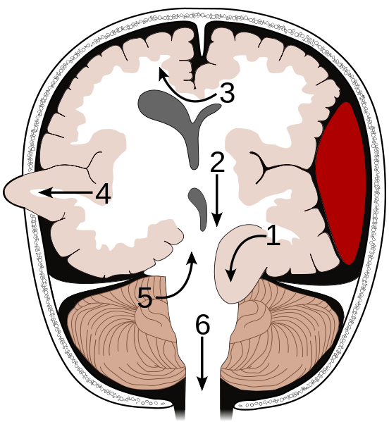
1. ungulate (caudal transtentorial)
2. central (more involved caudal transtentorial)
3. subfalcine (cingular)
4. transcalvarial
5. rostral transtentorial
6. cerebellar coning