Mission
Neurology Networks tries to offer broad exposure to various topics that may be presented on the veterinary neurology board exam.
MRI 4
Q.
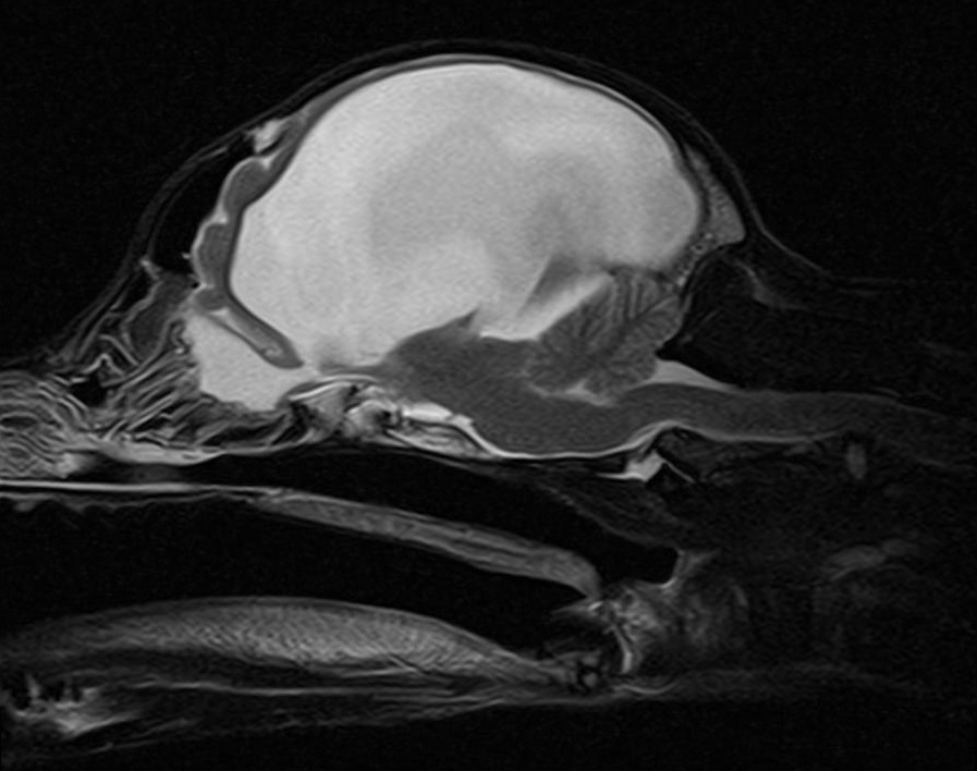

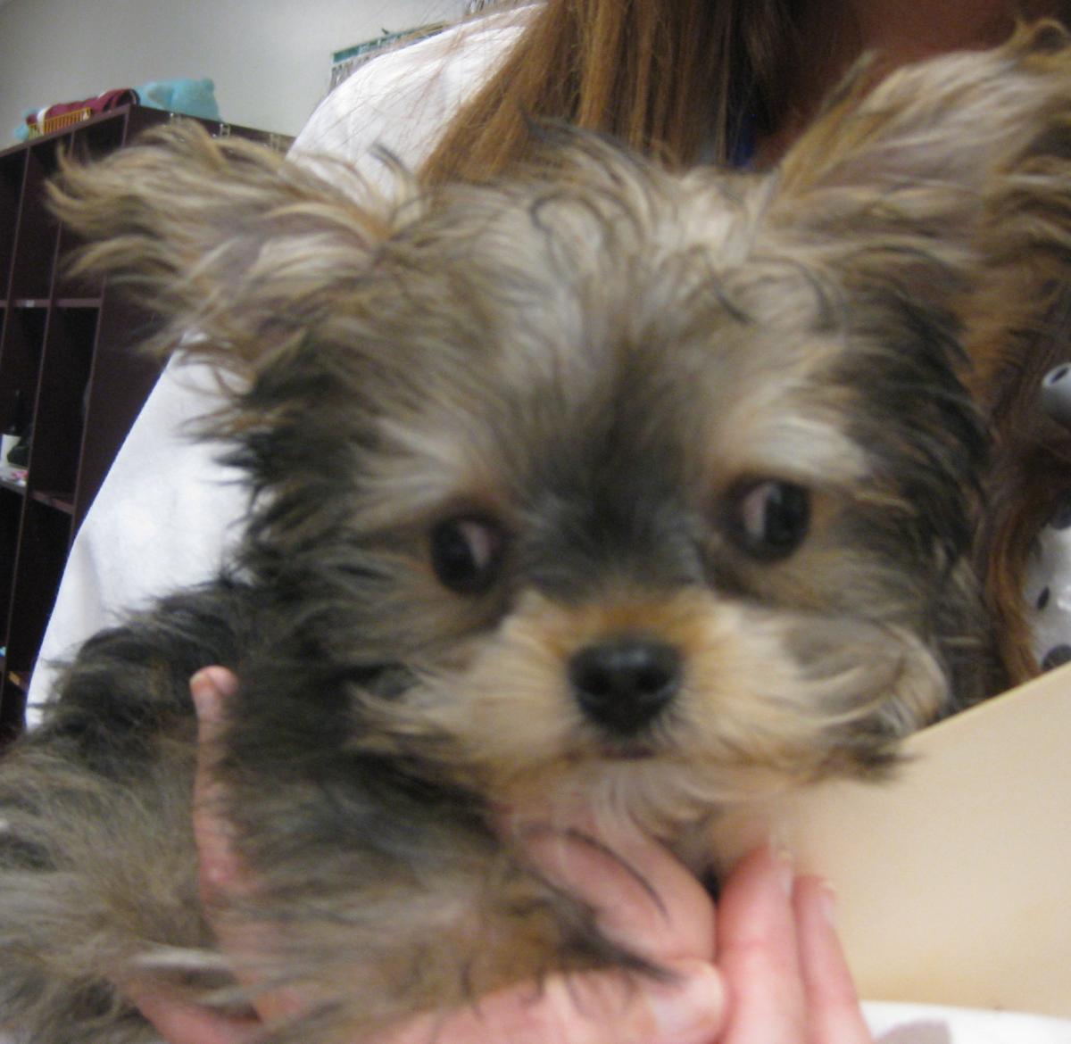
These are MRI images from a 10-month-old male intact Yorkshire terrier with a history of recent onset of lethargy. The dog is friendly and interactive but does appear quieter than normal for a dog of this age. You notice the facial features depicted in the photo below. He has poor menace OU with intact visual tracking, occasional subtle intentional head tremor, and slow conscious proprioception in all four limbs.
The owners have come to you for a second opinion regarding treatment options for this condition. What do you tell them?
A. This dog has severe hydrocephalus that is likely congenital. In these cases, fluid accumulation is thought to be associated with congenital microstructral anomalies that affect overall CSF flow and proper drainage. There are several clinical features seen in this dog including the ventrolateral strabismus (“setting sun sign”), abnormal mentation, domed head, and overall unthrifty appearance. The MRI confirms a severe form of the structural disease. Fortunately the changes are slow which allows for accommodation over time, allowing these dogs to maintain good overall function. However, because there is so little normal cortical brain tissue left, these dogs are also at great risk for slowly progressive worsening of signs with time or acutely with minor trauma or other encephalopathies. The lethargy in this case may be associated with progressing hydrocephalus, especially if no other underlying disease can be appreciated on systemic testing.
Treatment options can be medical or surgical. Medical management is meant to help treat acute deterioration, delay surgery, or if surgery cannot be performed or is inadvisable. Medical management focuses on:
Reducing CSF production
Managing periventricular edema/ brain swelling
Controlling any additional signs (ie seizures)
The more benign drug to use for reducing CSF production is omeprazole. Other options can include acetazolamide, furosemide, and prednisone (furosemide and prednisone do reduce CSF production but may help with edema and oevrall fluid pressure). All of these come with a higher risk of side-effects, especially if used chronically.
Surgical management is meant to be used in dogs early in the condition to help improve (or at least stabilize) signs and prevent further loss of brain tissue. Surgery most commonly involves placing a shunt to help drain excess fluid from the brain to the abdomen. This procedure is considered a definitive treatment, however it does carry a relatively high risk of complications, particularly in a dog with such severe hydrocephalus. This can include shunt obstruction, migration, overshunting with hemorrhage and cortical injury, and infection. These complications can warrant revision, removal, or can lead to permanent clinical worsening of signs or even death. Additionally the shunt is costly with prices for the shunt tubing alone costing hundreds of dollars or more.
In this dog’s case, it is reasonable to start with medical management. This will involve omeprazole 1mg/kg every 24 hours initially. Prednisone can be added if his signs are not yet improving. If his signs continue to deteriorate, then a shunt can be considered in an additional effort to better stabilize his signs.
“Hydrocephalus in dogs and cats”
William Thomas
VCNA Small Animal Practice. 40; 2010: 143-159
“Outcome of ventriculoperitoneal shunt implantation for treatment of congenital internal hydrocephalus in dogs and cats: 36 cases (2001-2009).”
Miriam Biel1; Martin Kramer; Franck Forterre; Konrad Jurina; Oliver Lautersack; Klaus Failing; Martin J Schmidt
J Am Vet Med Assoc. April 1, 2013; 242(7):948-58.