Mission
Neurology Networks tries to offer broad exposure to various topics that may be presented on the veterinary neurology board exam.
CT 2
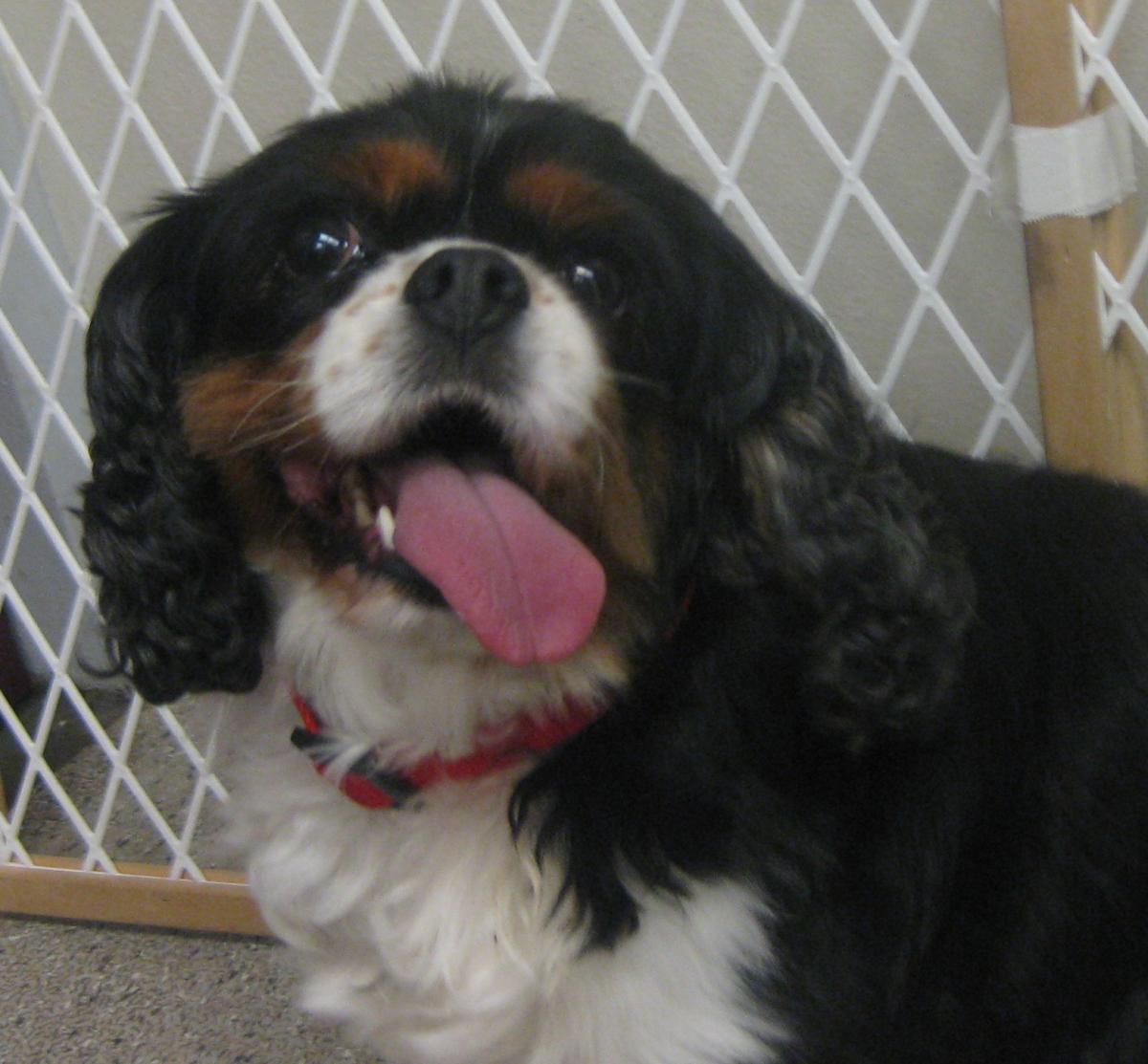
Q. This image is taken from a 4 year old female intact Cavalier King Charles Spaniel. This dog was diagnosed at a 2.5 years of age with caudal occipital malformation syndrome (COMS) and syringohydromyelia (SM): asymptomatic, grade 2 cerebellar impingement, grade 2c syringohydromyelia. In the last 2 weeks, she has developed the signs seen in the photo below.
Interpret this CT image:
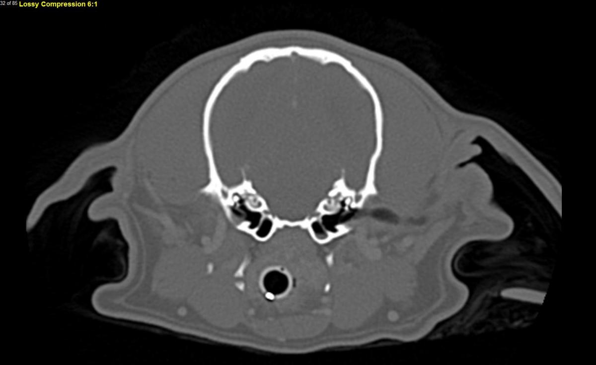
A. This is an axial post-contrast computed tomography image of the canine skull taken at the level of the tympanic bullae. There is a small amount of soft tissue opacity material in the ventral aspect of the left tympanic bulla and a possible slight defect of the tympanic membrane at the dorsal aspect on that same side. There is not overt lysis or other changes in the bony structures. The primary differential is infectious otitis media, though early primary secretory otitis media, injury after ear cleaning, neoplasm, and central brain lesion cannot be fully ruled out. Myringotomy with cytology and cultures is recommended following the CT scan to try to confirm infection.